
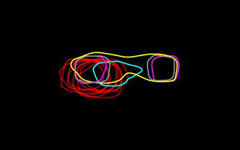
A comparison of the outlines of the super-resolved, aligned and averaged intensity distributions for different SPB pore membrane proteins caught in the act of duplication. Credit: Sue Jaspersen, Zulin Yu and Jay Unruh, Stowers Institute for Medical Research
A new imaging technique from researchers at the Stowers Institute for Medical Research and the University of Colorado, Boulder (USA), catches spindle pole body (SPB) activity in live yeast cells prior to mitosis—the process of cell division (eLife, doi: 10.7554/eLife.08586). The scientists believe that the technique, dubbed two-colored structured-illumination microscopy with single particle averaging (SIM-SPA), not only can illuminate a key step in the cell cycles of both yeast and higher animals, but could also record other tough-to-capture intracellular processes.
SPBs are cell bodies that divide before mitosis and that play a role in organizing the microtubules, internal cellular structures that help pull chromosome pairs apart and ensure that each daughter cell has a full set. Current optical microscopy methods can be used on living cells, but they cannot pick up these nanoscale structures; electron microscopy techniques, while capable of capturing the 200-nm SPBs, can only be used with fixed, nonliving cells. Duplication of yeast SPBs prior to cell mitosis is thought to occur over many discrete steps, ending with the duplicate SPB’s insertion into the nuclear envelope (the membrane surrounding the nucleus). However, these steps are not well understood.
To get a better look, the researchers labeled each of the 18 SPB components before duplication with endogenously expressed fluorescent protein derivatives. SIM creates a laser-generated pattern of horizontal lines on the SPB. By analyzing how the SPB interferes with the patterns, the researchers were able to image the structure and increase resolution. To help the team sort through all the “noise” created during SIM, the researchers used a modified SPA technique that employs 3-D image fitting of protein intensity distributions to improve image alignment (in the range of 10 to 30 nm) and to assist with quantification of color intensity.
During the team’s imaging demonstrations, SIM-SPA’s high resolution and quantitative fluorescence revealed that SPB duplication is initiated by the formation of an asymmetric filament, followed by the assembly of two other complexes. This assembly occurred at the same time the duplicate SPB is inserted into the nuclear membrane. And, surprisingly, SPB duplication occurred close to the end of mitosis rather than near the beginning. Since SPBs share a number of components with centrosomes, their animal counterparts, an improved understanding of SPB function and activity in yeast cells could also shed light on human cell division.