
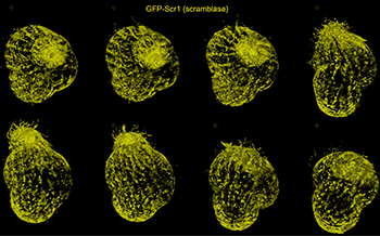
Volume renderings at eight consecutive time points of a single specimen of the protozoan T. thermophila taken from a 4-D data set. [Credit: Chen et al., Science 346, 1257998-1 (2014).]
Real time in vivo 3-D imaging is a valuable tool for understanding subcellular biological activity. However, until recently, available technologies forced scientists to make compromises between spatial resolution, imaging speed and level of phototoxicity to the specimen. Now, a group of scientists led by 2014 Nobel Laureate Eric Betzig has reportedly improved upon light-sheet microscopy to address these limitations with ultrathin light sheets created from 2-D optical lattices (Science, doi:10.1126/science.1257998).
Light-sheet microscopy employs an excitation lens perpendicular to the widefield detection lens in a fluorescence microscope to “section” a subject, illuminating only a single focal plane rather than its entire thickness. The light sheet is swept through the specimen to image a series of focal planes, which are then compiled to create a complete 3-D image. This technique has proven effective at the cellular level, but most light sheets created with Gaussian beams are too thick for subcellular imaging.
To get down to the subcellular level, Betzig and the team used non-diffracting Bessel beams, which they split into seven parallel parts to reduce the imaging time for capturing dynamic processes. This had the added benefit of decreasing light-induced damage to the cell, or phototoxicity, by spreading the light energy over a broader area, and illuminating only one plane at a time rather than the entire subject.
Making the imaging even less invasive, as well as sharper and faster, required making the light sheets ultrathin. For that, the team turned to non-diffracting 2-D optical lattices—periodic interference patterns formed by the superposition of plane waves traveling in well-defined directions. These new light sheets spread the light intensity even further and reduce the instantaneous power put on any given area of the cell, keeping the specimen healthier. The technique also improves axial resolution, as the interference pattern eliminates some undesirable light, and the thinness of the sheet increases imaging speed.
The group demonstrated their method on 20 different biological processes, including the diffusion of single transcription molecules in stem cell spheroids and embryonic development in nematodes and fruit flies. These improvements could allow microscopes to image and reveal new information about a wider array of biological processes.