
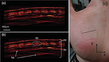
Top left: Axial slice of the reconstructed data acquired from the palm. Bottom left: Imaged skin: epidermis (ep), dermis (de), dermal papillae (dp) and horizontal plexus (hp). Right: The subject's palm, with the dotted line representing the slice of data shown in the first two images. Credit: Technische Universität München and Helmholtz Zentrum München
To diagnose skin diseases, physicians would like to have a reliable, non-invasive imaging technique to replace the slow process of examining biopsied tissue under the microscope. However, living skin severely scatters light at optical frequencies, prohibiting conventional imaging beyond a depth of a few hundred microns.
Scientists in Munich, Germany, have developed an optoacoustic technique that reaches several millimeters deep to reveal the underlying vascular structure of skin (Opt. Lett., doi:10.1364/OL.39.006297). The optoacoustic mesoscopy can be adjusted to show the tiny blood vessels near the junction of the epidermis and dermis or larger vessels deeper in the skin tissue.
Optoacoustic (or photoacoustic) imaging involves sending laser pulses into biological tissues, where some of the light energy is converted into heat, briefly causing thermoelastic expansion that in turn generates acoustic waves at ultrasonic frequencies. The team from the Institute for Biological and Medical Imaging (IBMI) used a 532-nm pulsed laser for their recent experiments.
Other teams have been experimenting with optoacoustic imaging, but the IBMI group achieved its results by using a custom-made ultrawideband lithium niobate transducer with a frequency spectrum ranging from a few millihertz to 200 MHz, with a central frequency of 102.8 MHz. The researchers wanted to see if they could detect smaller blood vessels, up to 25 μm across, that were invisible at smaller bandwidths with lower central frequencies.
The researchers imaged a 5-mm-wide square of skin on the palm of a healthy man's hand. A specially designed water-filled glass and polymer tank sat between the skin's surface and the acquisition equipment to acoustically couple the skin with the transducer. Computer processing of the signals showed the blood vessels of the dermis and even some vessels below the dermis.
In future work, the IBMI scientists will use near-infrared lasers to try to image the skin at greater depths, and they will redesign the equipment for potential clinical use.
IBMI is a joint institute of the Technical University of Munich and Helmholtz Zentrum München, Germany's environmental health research center.