
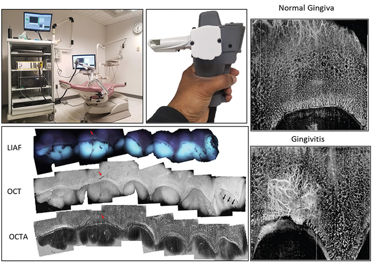
 Top left: Intraoral multimodal handheld OCT system in dental-office setting. Top center: Close-up of handheld imaging probe. Bottom left: The teeth and gingiva of the top-right palatal quadrant as imaged by the new imaging probe, including montaged images of raw LIAF, OCT en face structures, and OCT-A en face angiography of the teeth and gingiva. All en face images are projected by maximum intensity. Red arrow: gingival scar; black arrows: enamel crack. Right: Angiographic maps of normal gingiva (top) and gingivitis (bottom). [Enlarge figure]
In recent years, dentistry has witnessed tremendous advances in various radiographic-imaging modalities, including X-ray computed tomography (CT), cone-beam CT, intraoral periapical X-rays and 3D optical scanners for orthodontic CAD/CAM applications. Even with these advances, however, an unmet need remains for a noninvasive, nonionizing, high-resolution depth-resolved imaging modality for early detection and diagnosis of oral conditions in both soft and hard tissues.
Optical coherence tomography (OCT) is a very powerful imaging modality that meets those criteria and has expanded rapidly in areas outside of dentistry, including ophthalmology, endoscopy and dermatology. For dental applications, there is increased interest in using OCT for the investigation of oral diseases, including mucosal abnormalities, periodontal diseases and dental caries (tooth decay). The design of current commercial and non-commercial OCT systems, however, is not sufficiently flexible to accommodate intraoral, full-mouth inspections; as a result, the scan region is typically limited to the buccal (outside-facing) side of anterior teeth. A need remains for handheld or endoscopic OCT probes applicable for intraoral use in the oral cavity.
To help meet the need for OCT suitable for intraoral multimodal imaging, we combined OCT and OCT angiography (OCT-A) with light-induced autofluorescence (LIAF) into an intraoral compact handheld probe.1 The OCT system is based on a 1310-nm swept-source laser running at a line rate of 200 kHz and a bandwidth of 100 nm, with a Michelson interferometer setup.
Like OCT, LIAF is also a nonionizing imaging technique that focuses on the autofluorescence and absorption properties of the teeth and biofilm as caries are formed. In our setup, LIAF is excited by 395-nm (violet) light and detected by a visible-spectrum CCD camera.
We believe the system is suitable for routine intraoral clinical imaging applications, and capable of providing high-resolution, depth-resolved microstructural, angiographic and light-induced fluorescence images with a field of view of approximately 6.9×7.8 mm2 for diagnostic and monitoring applications.
Researchers
Hrebesh M. Subhash and LaTonya Kilpatrick-Liverman, Colgate Palmolive, Piscataway, NJ, USA
Nhan Le, Jie Lu, Peijun Tang, Kwok-Hung Chung and Ruikang K. Wang, University of Washington, Seattle, WA, USA
References
1. N. Le et al. Biomed. Opt. Express 13, 3629 (2022).