
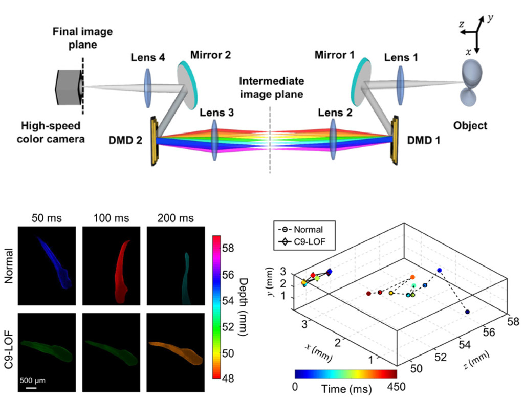
 Top: Schematic diagram of the DECALF system. Bottom left: Comparison of escaping behaviors of normal and C9-LOF zebrafish, showing all-focused frames at three time points. Bottom right: 3D traces after water-stream stimulation. [Adapted from J. Liu et al., Optica 8, 139 (2021)]
Light-field imaging can simultaneously capture 4D information of incident rays, allowing multiple-perspective viewing, digital refocusing and depth estimation. These advantages have spurred applications in diverse areas, from consumer photography1 to biomedical research.2 However, conventional light-field imaging based on microlens arrays involves an intrinsic trade-off between spatial and angular resolutions. While coded-aperture light-field (CALF) imaging can resolve that trade-off, existing CALF imaging systems—limited by the performance of aperture-encoding devices—are rarely used with broadband light and for dynamic events.
This year, we reported dispersion-eliminated CALF imaging (DECALF) that overcomes these limitations.3 DECALF is built on two interlinked 4-f imaging systems, in which two identical digital micromirror devices (DMDs) are placed on their Fourier planes. Leveraging the symmetry in the system design, the dispersion induced by the first DMD is fully compensated for by the second DMD.
This dispersion compensation allows DECALF to operate with light that covers the entire visible spectrum. A light-field dataset with full pixel counts is captured by sequentially opening each of the 5×5 square sub-apertures on the first DMD. The DMD’s high pattern refreshing rate has enabled DECALF to accomplish broadband light-field imaging at 20 fps.
We used DECALF to study the escaping behaviors of freely moving C9ORF72 loss-of-function (C9-LOF) zebrafish—a disease model developed to investigate the pathogenesis of the neurodegenerative disease amyotrophic lateral sclerosis. DECALF captured 3D movies that quantitatively tracked the locations and velocities of these C9-LOF zebrafish in the x, y and z directions after a water-stream stimulation was applied. The results revealed the slow responses and a limited moving ability of the C9-LOF zebrafish due to motor deficits, in contrast to the quick escape response behavior of normal counterparts.
DECALF imaging extends the operation scope of DMD-based CALF imaging to broadband light in real time. Its accommodation of the full visible spectrum not only enhances light throughput but also avoids the reduction of spatial resolution by pixel binning and the decrease in image quality due to laser speckles. Its video-rate 3D tracking ability could assist analyses of the dynamics of animals in response to various stimulations. And its compatibility with white light circumvents the potential color-induced complexity in the study of animal behaviors.
We thus envision that DECALF will be of general interest in the communities of optical engineering, imaging science and biophotonics.
Researchers
Jingdan Liu, Charlotte Zaouter, Xianglei Liu, Shunmoogum A. Patten and Jinyang Liang, Institut National de la Recherche Scientifique, Varennes, Quebec, Canada
References
1. R. Ng et al. Stanford Technical Report CTSR 2005-02 (2005).
2. R. Prevedel et al. Nat. Methods 11, 727 (2014).
3. J. Liu et al. Optica 8, 139 (2021).