
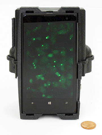
A smartphone-based device shows images of cells with genetic mutations. [Image: UCLA, Stockholm University and Uppsala University]
Integrating molecular analysis data with standard morphology-based pathology could improve the accuracy of healthcare providers’ diagnoses, thus allowing them to offer precise treatments based on the genetic makeup of a patient’s disease. Now, a team of researchers from Sweden and the United States has demonstrated a smartphone-based microscope that could deliver molecular diagnostic data directly to the healthcare provider, eliminating the need to send samples to outsourced laboratories—or, in the researchers’ words, take data “from the point of care to the point-of-expertise” (Nat. Commun., doi: 10.1038/ncomms13913).
OSA Fellow Aydogan Ozcan, of the University of California, Los Angeles, USA, and Mats Nilsson, of Uppsala University and Stockholm University, Sweden, led the team in designing and demonstrating the new telemedicine tool. They say results from their demonstration show that the smartphone-based multimodal microscope could facilitate the integration of targeted DNA sequencing and in situ point-mutation analyses with standard tissue morphology— without sacrificing accuracy—and at a fraction of the cost of traditional molecular diagnostics.
Design enabled by additive manufacturing
The researchers used 3-D printing technology to manufacture the relatively light optomechanical smartphone attachment, which has two compact laser diodes for multicolor fluorescence imaging and a white light-emitting diode for bright-field transmission imaging. It also has a 3-D stage that enables sample movement and alignment along the x, y and z axes.
The attachment’s external lens module has a focal length of 2.6 mm, which gives the smartphone camera a half-pitch resolution of 0.98 µm and an imaging field-of-view of about 0.8 mm2.
Molecular assays using smartphone microscopy
To test the microscope’s ability to detect individual molecules, the researchers developed a targeted sequence library of rolling-circle amplification (RCA) molecules with fluorescently labeled DNA repeats. (An RCA library is a traditional tool for DNA detection.) They found that the microscope could indeed image and sequence individual RCA molecules in solution.
The team conducted a similar sample prep and sequencing scheme demonstration using DNA extracted from three colon-cancer biopsies with fluorescently labeled oncogenes. Results from this demonstration were consistent with results from a standard benchtop fluorescence microscopy and sequencing analysis.
The researchers’ final demonstration tested the smartphone microscope’s clinical applicability to detect oncogene mutations in cancer cells—directly in colon cancer tissue in situ. Their data showed that the smartphone microscope achieved 100 percent concordance to a standard clinical genotype analysis and whole-tissue scanning with an automated benchtop fluorescence microscope.
Ozcan has said that the smartphone-based microscope could cost as little as US$500 if it were produced in high volume, making it potentially attractive to field and limited-resource settings. (For reference, the standard microscope used to validate the smartphone version would cost about US$50,000).