
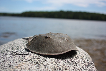
A horseshoe crab in profile, ready for its close-up. One of its compound eyes is visible. [Image: Pixabay/ckaras]
The compound eyes of the horseshoe crab—an arthropod whose evolutionary roots stretch back 480 million years or more—have long intrigued scientists. And, while they have learned a great deal about the structures of the critter’s eyes, researchers continue to debate just how those structures work to execute their primary function: concentrating and focusing light.
A multinational research team has now used multiple optical and other techniques to dive into the question (Adv. Sci., doi: 10.1002/advs.202203371). Their work suggests that evolution has made the most of the capabilities of the horseshoe crab’s exoskeleton, or cuticle, to build compound eyes of remarkable complexity and subtlety. Quite apart from the biological interest, the team thinks the work could advance bio-inspired design of more sustainable optical materials and components.
Ten-eyed wonder
Notwithstanding its name, the horseshoe crab (Limulus polyphemus) is not a crab at all, but a member of the arthropod subphylum known as the chelicerates—the same subphylum that includes arachnids, or spiders. And, like their spider cousins, the horseshoe crab is covered by an exoskeleton, or cuticle, consisting of the natural polymer known as chitin, in a composite structure with other proteins.
One of the uses to which nature has put that chitin exoskeleton is in forming of the creature’s ten eyes. These include six primitive eyes sensitive to light-dark cycles, and distributed in various parts of the horseshoe crab’s body; two ocelli that are sensitive to UV light; and two lateral compound eyes. This last pair—each of which includes around a thousand substructures, or ommatidia—is, neurophysiologically speaking, where the heavy lifting of the horseshoe crab’s vision takes place.
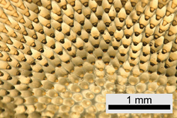
A transmission light micrograph of the inside of the compound eye of the horseshoe crab, showing its conical, light-collecting structures. [Image: Oliver Spaeker]
GRIN or TIR?
Just how the compound eye’s ommatidia—which each consist of a cornea, a conical lens-like structure and a cluster of photoreceptors—actually work in the horseshoe crab to gather light and guide it to the photoreceptors has proved the source of some disagreement.
One line of thinking has focused on the gradient of refractive indices across the conical lens structure. That suggests that the conical structure could function as a refractive focuser, using the same principles a graded-index (GRIN) optic or lens. But other researchers have emphasized the overall shape of the conical light guide, and the potential role of total internal reflection (TIR) at the interface between the cornea and the surrounding tissue in collecting and channeling the light.
The team behind the new research, led by Yael Politi of the Center for Molecular Bioengineering at Technische Universität Dresden (TU Dresden), Germany, used a variety of complementary techniques to enrich the data and modeling of the horseshoe crab’s eye, in the hope of distinguishing between the GRIN and TIR models. As a first step, they used X-ray diffraction, or XRD (conducted at the European Synchrotron Radiation Facility in Grenoble, France, and the BESSY II Light Source in Berlin, Germany), to map the structural and compositional variation of the eye components at multiple length scales. This gave an overall view of how evolution has used the materials at hand—mainly chitin, other proteins, and water—in different proportions and cross-linked structures to vary the optical properties across the cones.
Micro-Raman spectroscopy sharpened the structural picture from the XRD. And X-ray fluorescence revealed that, in certain areas, the cuticle material is doped with element bromine—known to act as an exoskeleton-stiffener in insects, but also possibly related to achieving locally high refractive-index gradients.
Modeling the compound eye
Next, the team used quantitative phase imaging (QPI) to measure the phase shift of transmitted light, and thereby carefully map the refractive-index distribution across the cornea both longitudinally and in cross-section. That let the researchers tightly correlate the structural and compositional changes with consequent changes in optical properties. Finally, the team used full-wave optical modeling and ray-tracing simulations to gain insight on how the mapped refractive-index profiles and the shape of the cones might contribute to guiding incident light from the cornea to the photoreceptors.
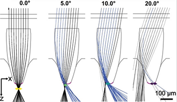
Ray-tracing simulation results showed how the importance of total internal reflection along the corneal interface (blue rays) increasingly affects light guidance as the incidence angle increases. [Image: O. Spaeker et al., Adv. Sci., doi: 10.1002/advs.202203371 (2022); CC-BY 4.0]
The team’s modeling suggested that both the GRIN and TIR models are at work in the eyes—under different circumstances. At normal, straight-on incidence angles, GRIN-based refractive mechanisms predominate in focusing the light. But at higher incidence angles, total internal reflection in the cornea starts to become important, and a combination of TIR and GRIN mechanisms together serve to guide the light to the receptors.
Learning from nature
The research team believes that evolution’s use of chitin, proteins, water and a dash of bromine to create the subtle and hierarchical optics in the horseshoe crab’s compound eye offers “a fascinating example of the flexibility and multifunctionality of arthropod cuticle, based on a simple ‘blueprint’ and made from modular building blocks.” Indeed, in a press release accompanying the research, team leader Politi said that “using cuticle to build lenses is unusual and the extent to which the horseshoe crab repurposes it for its eyes is nothing short of remarkable.”
Politi believes that optical and materials designers can learn from the horseshoe crab’s example. “The compound eye is nature’s answer to the need of capturing a wide field of view in an otherwise small eye,” she said. “It’s a similar challenge to that faced by modern camera designers who want to further and further miniaturize the cameras while keeping a wide field of view of above 90 degrees.” The team hopes that, in the long run, studies such as this one can provide new tools and insights for developing bio-inspired materials and devices.
In addition to contributors from TU Dresden and the European Synchrotron Radiation Facility, the work also included researchers from the Max Planck Institute of Colloids and Interfaces, Germany; the Institute for Globally Distributed Open Research and Education, Brazil; the University of Salzburg, Austria; the University of Fribourg, Switzerland; and the firm Telight, Czech Republic.