
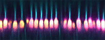
Using a modified confocal microscope, researchers observed the optical properties of mitochondria in living cone photoreceptors from mammalian eyes. The team found that the mitochondria acted as microlenses that concentrated light, facilitating its transmission from the inner to the outer segments of cone photoreceptors. [Image: J. Ball, NEI]
The retinas of humans and primates have long held a mystery. In these mammals, immediately next to the light-sensitive outer segment of the retina’s cone photoreceptors, sit tightly packed bundles of mitochondria—the organelles that function as the cell’s energy factories. Scientists have assumed that the odd position of these cellular powerhouses was mainly to allow efficient energy delivery to the outer segment. But increasing evidence has suggested other pathways for this energy transfer.
Now, after studying photoreceptors of ground squirrels using a modified confocal microscope, researchers in the United States revealed another possible role for the retina’s mitochondrial bundles: as tiny lenses that focus light onto the photoreceptors’ outer segments (Sci. Adv., doi: 10.1126/sciadv.abn2070). The researchers write that the newly found optical function of mitochondria could also have “imperative clinical implications,” particularly in aiding retinal disease diagnosis.
Bundle of mystery
Mitochondria are membrane-bound organelles (essentially internal organs of eukaryotic cells) that generate the cell’s energy. For most such cells, these organelles form networks surrounding the cell nucleus. In the retinal photoreceptors (particularly the cones) of some mammalian species, however, mitochondria also occur as tightly packed and elongated bundles.
This has perplexed scientists for some time, as the dense geometry of mitochondria might be expected to scatter the light before it reaches the photoreceptors’ outer segment. But the complexities of separating the mitochondria bundle’s optical properties from those of the rest of the retina have held back progress on solving the puzzle.
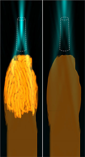
Left: Computer simulations by the team suggested that when the cell membrane and cytoplasm are paired with bundles of mitochondria (bright yellow), the light (blue) is concentrated on the outer segment (white dashed line). Right: In simulations without the mitochondria, the light did not focus on outer segment. [Image: J.M. Ball et al., Sci. Adv. 8, eabn2070 (2022); CC BY 4.0]
To tackle this mystery, three researchers at the National Eye Institute, a part of US National Institutes of Health, decided to study thirteen-lined ground squirrels (Ictidomys tridecemlineatus), whose retinas mostly comprise cone photoreceptors. The researchers first isolated the photoreceptor layers from the squirrels’ eyes. They then observed the live photoreceptor samples using a confocal microscope, with the condenser replaced by a dichroic mirror to direct collimated LED light in the same way it passes through the retina.
The researchers discovered that instead of scattering the light, the mitochondria bundles focus light it in a thin, pencil-like trajectory, concentrating it onto the cone photoreceptors’ outer segments for easy detection. The finding matched up with separate computational modeling by the team.
Clinical and evolutionary implications
“We were surprised by this fascinating phenomenon that mitochondria appear to have a dual purpose: their well-established metabolic role producing energy, as well as this optical effect,” said Wei Li of the National Eye Institute, in the press release accompanying the study. At the same time, “the lens-like function of mitochondria also may explain the phenomenon known as the Stiles–Crawford effect,” said John Ball, also of the National Eye Institute.
The Stiles–Crawford effect is a phenomenon where the light entering through the edge of the pupil elicits a lower photoreceptor response than the light of the same intensity entering near the pupil’s center. This angular dependence of light, the researchers note, mirrors the pathway the light takes after being directed by the mitochondrial microlenses.
The researchers think connecting the two phenomena could have significant clinical implications. Mitochondrial dysfunction was long thought to be the origin of many retinal degenerations. The new findings on the mitochondria’s dual use could encourage broader application of the Stiles–Crawford effect for noninvasively diagnosing those disorders.
The team also points out that the newly found role of mitochondria bundles resembles that of the oil-droplet microlenses found in the eyes of many birds and reptiles. Indeed, said Li, “this insight conceptually bridges compound eyes in arthropods with the camera eyes of vertebrates, two independently evolved image-forming systems, demonstrating the power of convergent evolution.”