
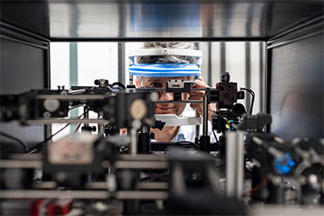
Researchers developed a two-photon excited fluorescence scanning laser ophthalmoscope that can be safely deployed to image the visual cycle. [Image: International Centre for Translational Eye Research - ICTER, Grzegorz Krzyzewski]
Ophthalmoscopy, performed as part of a routine eye exam, images the back of the eyeball, called the fundus, to check for symptoms of retinal detachment or eye diseases such as glaucoma. In recent years, advancements in optical techniques have led to much more detailed structural information compared with traditional fundus photography.
Now, researchers in Poland and the United States have developed a two-photon excited fluorescence scanning laser ophthalmoscope that probes not just the structure but the biochemistry of vision (J. Clin. Investig., doi: 10.1172/JCI154218). The instrument non-invasively measures metabolic processes occurring in the human retina, potentially allowing for earlier and more accurate disease diagnosis.
Imaging the visual cycle
Current optical methods for ophthalmoscopy provide high-resolution images of the retina and other structures. However, they can only uncover abnormalities after disease is already present in a patient’s eye.
Maciej Wojtkowski and his colleagues aimed to create a new instrument and measurement method capable of detecting early age- or disease-related changes in the retina. Back in 2001, they pioneered the development of Fourier-domain optical coherence tomography (OCT) for in vivo retinal imaging. Now, the researchers wanted to observe the production of retinol, a photoactive molecule that triggers the vision process, as a measure of testing the efficiency of photoreceptors in the eye.
“Non-invasive in vivo imaging of this process, which we call the visual cycle, is a dream for vision scientists, but also for ophthalmologists and pharmacologists who could make faster diagnoses and monitor the effectiveness of therapies,” said Wojtkowski, of the International Centre for Translational Eye Research (ICTER) at the Polish Academy of Sciences.
Testing for safety
Biochemical markers like retinol must be excited with UV rays, which isn’t possible in practice due to the danger of damaging the cornea and lens of the eye. Instead, Wojtkowski and his colleagues decided to use a nonlinear process with two-photon excitation, which illuminates the eye with short pulses of near-infrared light (730–760 nm).
“For many experts using fluorescence in their work, it is obvious that two-photon excited fluorescence occurs on the retina,” he said. “However, what has not been obvious until now is whether it is safe for the eye.”
For their two-photon excited fluorescence scanning laser ophthalmoscope, the researchers first chose the length and the repetition time of a femtosecond laser pulse such that the light delivered to the eye would have a power below standard safety thresholds. Then, they confirmed that no damage to a mouse retina occurred from the instrument, followed by a similar experiment on Wojtkowski’s own eye.
Aiming for the clinic
While the signals obtained are weak, the researchers were able to use algorithms in combination with data from a traditional scanning laser ophthalmoscope to boost the image quality. A comparison with imaging results from mouse models of various visual disorders confirmed that their device can feasibly observe biomarkers actively involved in the visual cycle.
“As next steps, we need to identify and validate specific quantitative parameters that will become biomarkers used in clinical practice in the future,” said Wojtkowski. “To do this, we will also need to test multiple methods for extracting spectral information from the resulting fluorescence signals and perform many measurements on patients.”