
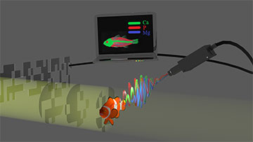
Researchers at Bar Ilan University, Israel, have combined computational ghost imaging with X-ray fluorescence measurement. The new method could produce chemical element maps in a high-resolution and efficient way. It could be useful for a range of applications in biomedicine, materials science, archaeology, art and industry. [Image: Sharon Shwartz, Bar Ilan University]
X-ray fluorescence (XRF) can tell scientists much about the chemical makeup of biological tissue, industrial machine parts and many other composite materials. However, X-rays are difficult to focus, and the raster scanning required to locate spatial variations in chemical composition is a painfully slow technique.
Now researchers at an Israeli university have sped up the XRF chemical mapping process by incorporating computational ghost imaging to create a nondestructive, lens-free detection technique (Optica, doi:10.1364.OPTICA.441682). Compressed sensing algorithms make up for the reduced number of measurement points compared to the usual XRF methods.
The proof-of-concept experiment yielded maps with a resolution of about 35 μm, although the research group could easily take that down to 5 μm or less, says Sharon Shwartz, a physicist at Bar Ilan University, Israel.
Ghostly imaging
Computational ghost imaging, first proposed in 2008, harnesses data processing to correlate two light beams that individually yield no information about the subject. Only one of the two beams interacts with the subject; the other carries a reference pattern.
The Bar Ilan team first measured X-ray beam intensity fluctuations created by a pixelated mask with features about 40 μm across. Next, the researchers put the target object—which contained separate small chunks of iron, cobalt and brass—into the beam and recorded the fluorescence emitted by the different metals. Artificial intelligence algorithms reconstructed the images for each emission line coming from the metals.
The key to success
According to Shwartz, other teams have previously demonstrated ghost imaging approaches in the X-ray regime, but only for transmission measurements. “The main novelty of the present work is the application of the computational ghost imaging approach for chemical element mapping,” she says. “It relies on the same technology that enables the [conventional] X-ray ghost imaging and, to the large extent, also on the same developments in algorithms [either compressive sensing or machine learning] and computer power that enable other computational imaging approaches with visible light.”
Ghost imaging in the visible range has to compete with traditional microscope technology, Shwartz says, but lensless computational methods with X-rays have great potential for improving the efficiency of the data collection process.
The key to the success of the experiment, Shwartz says, was modulating the intensity with a mask that induced intensity fluctuations that were higher than the noise. “We used simulations and found the modulation simply by measuring the intensity fluctuation by a camera,” she adds.
Future direction
Getting the technique’s resolution down to a few microns is easy with X-rays, according to Shwartz. Although electron beams could attain much higher resolution, the required high vacuum for electron-based systems would make their use impractical in medical settings. However, the microelectronics industry might someday use ghost imaging based on electron beams to test the next generation of chips.
Shwartz says her team has already begun to extend the method to three dimensions by using tomography to image the 3D structures of objects. “This will be a first step in the direction of using the method for a new modality for computed tomography [for medical applications],” she adds.