
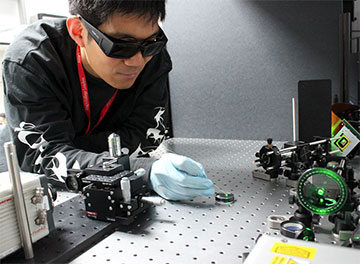
Researchers have created a photoacoustic imaging endoscope probe that can fit inside a medical needle with an inner diameter of just 0.6 mm. Tianrui Zhao, first author of the paper describing the work, is shown holding the imaging probe. [Image: T. Zhao, King’s College London]
Scientists at two British universities have developed an ultrathin photoacoustic endoscope probe that is two orders of magnitude faster than previous probes based on optical fibers (Biomed. Opt. Express, doi: 10.1364/BOE.463057).
The new device eliminates issues that plagued previous fiber endoscopes, such as bulky components and low imaging speeds. The probe fits inside a medical needle with an inner diameter of 0.6 mm, making it a potential aid for small-tumor biopsies and minimally invasive surgeries.
Types of endoscopic probes
Over the years, biomedical scientists have developed several types of probes to help them characterize abnormal tissues in situ. However, each probe had its limitations. Endoscopic optical coherence tomography gives surgeons 3D views of cells, but with low contrast. Fluorescence probes tag abnormal cells with fluorescent labels, but don’t provide 3D imaging.
Photoacoustic endoscopy was conceived as a best-of-both-worlds platform for both structural and molecular characterization of potentially dangerous cells. Originally, photoacoustic endoscopes contained a bundle of optical fibers, but in the last decade, researchers have replaced the bundle with a single multimode fiber. However, such scopes took as long as 30 seconds to acquire a single frame of image data because of the required liquid-crystal spatial light modulator for wavefront shaping.
Speeding things up
In their recent set of experiments, researchers at King’s College London and University College London replaced the spatial light modulator with an off-the-shelf digital micromirror device. The group wrote up a fast and efficient algorithm to control the micromirror, which was the key to success, according to Wenfeng Xia, a biomedical engineer at King’s College London and leader of the research team.
In the experimental setup, the digital micromirror shaped light from a 532-nm laser into focused pulses and passed the light into a multimode fiber, which was inserted into a 20-gauge medical needle. At the tip of the hollow needle, the light pulses hit the biological structures of interest, such as red blood cells, which then generated acoustic waves. An ultrasound sensor based on a plano-concave microresonator, located at the tip of a single-mode fiber also inserted into the needle, detected the acoustic signal, and the single-mode fiber carried the signal for image processing.
For a proof of concept, the group acquired high-resolution images of red blood cells from a mouse blood sample. Researchers were able to cover a 100-μm-wide area at an imaging rate of 3 frames per second.
Future directions
In future work, Xia says, the researchers will use the endoscope on an in vivo animal model to see how well it can work in detecting small tumors. Xia’s group had already tested the graded-index multimode fiber to make sure it could resist modest bending during an endoscopic procedure, but more testing for robustness needs to be done. Finally, the device would have to gain regulatory approval before it could be used in patients, Xia says.
Correction, 6 September 2022, 10:30 EDT: The original version of this story said that a continuous-wave laser was used to supply light to the digital micromirror device; the actual laser used was a pulsed 532-nm laser.