
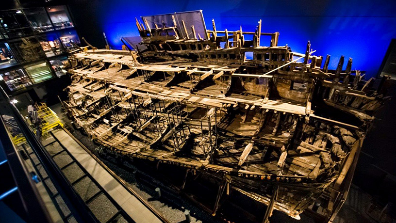
An international research team used an X-ray technique developed by Columbia University and the European Synchrotron Radiation Facility to identify zinc-containing nanoparticles lodged within the wooden hull of the Mary Rose, Henry VIII’s favorite warship. These nanoparticles are leading to deterioration of the remains of the ship, which sank in battle in 1545 and was salvaged in the 1980s. The Tudor-era ship and its collection of 10,000 artifacts are now housed in the Mary Rose Museum in Portsmouth, UK. [Image: Mary Rose Museum] [Enlarge image]
An X-ray technique developed to study catalysis and batteries for energy conversion and storage has a new application: archaeology and cultural artifacts.
Using the technique—known as computed-tomography PDF, or ctPDF—an international team of researchers has studied a Tutor-era warship that sunk in battle in 1545. The results, which revealed previously unseen nanoparticles, will help conservators preserve the ship from further decay (Matter, doi: 10.1016/j.matt.2021.09.026).
Preserving the past
After 437 years under the sea, the oak skeleton of the Tudor warship Mary Rose was salvaged from the English Channel in 1988. The ship, once King Henry VIII’s favorite, has been conserved in a museum of Portsmouth, UK, ever since but, despite conservation efforts, is slowly decaying.
To better conserve the Mary Rose, researchers need to know what’s going on in the wood at a molecular level. The new work achieves this by using an X-ray technique that combines methods of computed tomography and pair-distribution function (PDF) analysis to produce 2D and 1D images of a sample, which show both the position and nanostructure of inorganic materials within it. This method provides more information that other techniques, which might only give the distribution of chemical elements or the density of a material.
“What is special about ctPDF is it also tells you in quantitative detail the actual material that is there, and its structure. For example, in this study we found zinc sulfide nanoparticles, but could also measure the lattice constant, which is a measure of how close the atoms are to each other in the material,” said Simon Billinge, physicist at Brookhaven National Laboratory, professor at Columbia University and coauthor on the new study. “It really gives completely novel insights into what is going on in the artefact.”
Corrosive contaminants
The ctPDF analysis of the Mary Rose revealed zinc sulfide nanoparticles just 5 nm long. These particles were likely created by bacteria that lived in the wood of the warship when it was on the seafloor. Once removed from its anaerobic sea environment, that sulfide in the ship oxidizes into sulfate. In water, this creates corrosive sulfuric acid.
Now that researchers know about the zinc sulfide, they can develop strategies to better conserve the Mary Rose. Billinge expects the work will help other conservation studies as well, where it could be used in studying paintings, pottery and other materials.
“It is clearly a very powerful technique, combining quantitative nanostructure analysis with spatial resolution, that it will have many applications, especially as our tools for automating data acquisition and analysis get developed so that the experiments are more straightforward,” Billinge said.
The work was performed by researchers from Columbia University, USA, the European Synchrotron Radiation Facility (ESRF), the University of Sheffield, UK, the Mary Rose Trust, and the University of Copenhagen, Denmark.