
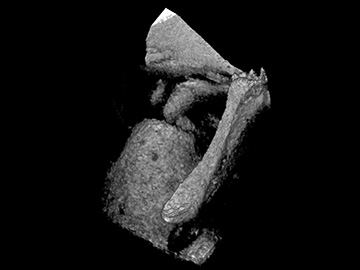
Optical coherence tomography (OCT)-generated image of a chinchilla middle ear. Researchers used an OCT vibrography tool to reconstruct the motion of ossicular bones in response to high-frequency sound. [Image: Wellman Center for Photomedicine / Massachusetts General Hospital]
A research team led by OSA Fellow Seok-Hyun Yun at the Wellman Center for Photomedicine at Massachusetts General Hospital, USA, reports using a form of optical coherence tomography (OCT) to capture 3-D anatomical and vibrational images of ossicles—tiny bones within the middle ear—as they move in response to sound (Biomed. Opt. Express, doi: 10.1364/BOE.9.005489). The imaging method, which they named OCT vibrography, uses OCT phase-synchronized with a range of high-frequency sound stimuli. In demonstrations with a chinchilla ear model, the researchers used OCT vibrography to observe a previously unknown mode of ossicular motion at high frequencies.
The new instrument, which is an improvement on the team’s previous design, may offer insights into how mechanical vibrations of ossicles contribute to sound perception in the brain. According to the team, it could someday also be used to diagnose hearing problems.
The role of the middle ear
When sound waves enter the ear, they stimulate the tympanic membrane (TM)—popularly known as the eardrum—and are picked up by a set of tiny bones called ossicles in the air-filled middle ear. The ossicles convert the sound waves into mechanical vibrations that are conducted to the fluid-filled inner ear where they are transformed into nerve signals that are sent to the brain.
Disruptions to the conduction of mechanical vibrations through the middle ear, either by disease or damage, can result in hearing loss. There are imaging methods available for viewing structures within the middle ear, but they are subject to artifacts caused by nearby vibrations or require surgical removal of the TM in order to access the structures beyond. OCT vibrography could, say the researchers, remove both of these barriers by using phase-synching as a way of only imaging the structures of interest (the moving ossicles) and using OCT as a way of imaging structures beyond the TM without surgical intervention. The technique may also shed light on exactly how sounds are conducted from the middle ear to the inner ear, which could greatly benefit hearing research.
New and improved OCT vibrography
This OCT vibrography system is actually an improvement on a previous design from Yun and his team. The new system offers more precise measurements of phase delays along the ossicles and can be used to measure acoustic transduction at frequencies above 10 kHz. (The previous model was limited to less than 3 kHz.)
After testing the system’s phase sensitivity and stability, the researchers used a chinchilla ear model to demonstrate the OCT vibrography tool because of its similarity to the human ear in size and sound-frequency sensitivity. First, they scanned the wavelength-swept OCT laser through the chinchilla’s auditory canal to get a 3-D image of the TM and ossicles in the middle ear. Next, they synchronized a high-frequency sound stimulus to the OCT measurement system. The sound caused the middle-ear ossicles to move, which triggered the OCT system to collect vibrography measurements.
The researchers completed several 60-second OCT vibrography scans that simultaneously measured the structure and movement of more than 10,000 points on the TM and ossicles over a range of sound frequencies. Using the volumetric data sets, they constructed 3-D “movies” of the ossicular motion with exaggerated vibration amplitude. Some of these movies showed a previously unknown mode of ossicular motion that meshed with theories about how high-frequency sound waves pass through the middle ear.
Future plans
Encouraged by the results of these initial demonstrations, the researchers hope to start testing OCT vibrography on human cadaver ears. However, Yun and his colleagues note that it will be a challenge to apply OCT vibrography to live subjects because heart beats and breathing during the 60-second scan can cause artifacts in the motion measurements. Therefore, they are working to see if they can get enough data to diagnose middle-ear problems from a rapid form of OCT vibrography that measures just three to five points on the ossicles and TM instead of the full set of 10,000 points.