![]()
A combination of an inward pull from optical gradient forces and a forward push from forward scattering creates an optical waveguide out of living bacteria. [Image: Courtesy of A. Bezryadina/SFSU]
Researchers using light to explore biological tissues always face the problem of scattering in opaque media—a problem that limits the light’s penetration depth. Now, work by a multinational research team suggests a novel solution: Use the living cells suspended in the medium to create an optical pipe that allows light to penetrate to greater depth (Phys. Rev. Lett., doi: 10.1103/PhysRevLett.119.058101).
The team—headed up by OSA Fellow Zhigang Chen of San Francisco State University (SFSU), USA, and Nankai University, China, and by OSA Fellow Roberto Morandotti of Université du Québec, Canada—shone a green laser at moderate power through a suspension containing a common strain of cyanobacteria. The researchers found that the microbes, driven by a combination of nonlinear optical effects, coalesced to form a needle-like waveguide that could transmit the light a distance of four centimeters through seawater, ordinarily a strongly scattering medium. What’s more, the bacteria survived their laser-light bath—an observation that, according the scientists, points to a variety of possible applications in biology and medicine.
From nanoparticles to cells
According to OSA Member Anna Bezryadina, a co-lead author on the new study (along with Tobias Hansson at Université du Québec), the idea of using cells to create a waveguide grew out of earlier work involving colloidal suspensions of nanoparticles. That work, led by Chen and by OSA Fellow Demetrios Christodoulides at the University of Central Florida, USA, had demonstrated that—with appropriate tuning of the refractive-index contrast between the particles and the surrounding medium—radiation pressure and optical gradient forces would cause the suspended particles to coalesce around an incident laser beam. The result was a sort of ad hoc optical fiber in the nanoparticle suspension, creating a “needle of light” that could propagate for several centimeters (Phys. Rev. Lett., doi: 10.1103/PhysRevLett.111.218302).

Anna Bezryadina.
The Chen team wanted to see if the same trick was possible in a suspension not of nanoparticles, but of biological cells. That would constitute a neat trick, as it would presumably open up “tremendous applications in medicine and biofabrication,” says Bezryadina. But biological cells seem at first glance like an unpromising candidate for making a waveguide: given that they’re mostly made of water themselves, the refractive-index contrast between them and the surrounding aqueous liquids they occupy isn’t particularly large. “When we tried it,” Bezryadina notes, “we were surprised at how well the idea worked.”
Blue-green bacterium meets green laser
To demo the idea, Bezryadina, Hansson and colleagues began with a seawater sample that contained a suspension of living Synechococcus bacteria—a cyanobacteria strain common in shallow marine waters (and, coincidentally, important in the global carbon cycle). They packed the suspension into a 4-cm-long tube, and then fired a 532-nm (green), 50-micron-wide linearly polarized laser lengthwise through the tube. (The wavelength choice was important: these blue-green bacteria have relatively low absorption at 532 nm, so the team could rule out thermal effects as an explanation of any observations.)
The team found that, in a seawater sample without the bacteria, the laser diffracted from 50 µm to around 650 µm, as expected. With the Synechococcus bacteria in the suspension, and at a low laser power (0.1 W), the linear scattering was even worse, with the beam expanding to a width of around 1.25 mm.
When the researchers pumped up the laser power to 3 W, however, the picture changed dramatically. Instead of scattering and spreading, the beam remained trapped in a needle of light around 200 µm wide for a substantial distance—with around 10 percent of the light in the needle propagating across the entire 4-cm length of the tube.
After the experiment, the team tested the survival of the cells in the suspension using standard viability assessment techniques. The researchers found that the mortality was only 0.1 percent higher in the laser-treated sample than in a control sample. Suspended in seawater, the optically nudged bacteria had formed a waveguide that transmitted laser light—and, so to speak, had lived to tell the tale.
Forward scattering
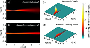
In a model that includes only the optical gradient force (top), the bacterial waveguide breaks up about halfway through the 4-cm experimental distance. A model that adds in forward scattering arising from radiation pressure (bottom) accounts for the deeper penetration of the guided light actually observed in the experiments. [Image: Phys. Rev. Lett., doi: 10.1103/PhysRevLett.119.058101] [Enlarge image]
It remained for the scientists to explain what was actually happening in the sample tube, a task for which they turned to numerical modeling. One obvious explanation was an optical gradient force; even with the small refractive-index contrast between the cells and the surrounding seawater, that force would be sufficient, at high enough laser power, to gather the cells around the beam path and create an effective waveguide.
However, the team’s modeling suggested that the optical gradient force, while necessary, wasn’t enough in itself to make the beam propagate for the full distance through the suspension. If only that force were acting, the beam would tend to self-focus and collapse about halfway through the full 4-cm length.
To get the extra distance observed in the experiments, the team needed to add a second effect to the model: forward light-scattering from the cells. The radiation pressure from that scattering, the modeling revealed, provided an extra optical “push” that moved the cells out in front of the propagating beam, extending the living-cell waveguide and allowing the thin optical “bio-fiber” to stretch across the lab tube’s entire 4-cm length.
A “new research direction”
In addition to experiments with Synechococcus, Bezryadina notes, the researchers also coaxed a variety of other cell types (including E. coli, red blood cells and a different strain of marine cyanobacteria) to form waveguides in a similar manner. The observation of the effect in other cells—and, more important, the fact that the cells can survive the ordeal of being temporarily marched into formation to serve as a conduit for laser light—opens up a variety of potential applications in the long haul, according to the team.
One such application, Bezryadina suggests, would be to enlist living cells to serve as light guides through scattering media, for imaging through biological fluids such as blood or amniotic fluid, or for “non-invasive medical diagnostics.” Another might be creating self-assembling, tunable biological microchips that include living-cell optical waveguides.
To get to these and other applications, though, will require a lot of additional work to account for different cell sizes, concentrations and types and to tune the optical parameters appropriately. “This is a very new research direction,” says Bezryadina. “We’ve demonstrated experimentally that light can penetrate deep into a biological environment without destroying it. That was something that, it was commonly believed, couldn’t be done, due to strong scattering and weak optical nonlinearity.”
In addition to researchers from SFSU, Nankai and Québec, the team included scientists from the University of Sussex, U.K., the University of Electronic Science and Technology of China, and the National Research University of Information Technologies, Mechanics and Optics, Russia.

