
Axial X-ray images of an infant mouse. From left: normal X-ray image, phase contrast image and dark field image. Credit: Elena Eggl / TUM
Today's computed tomography (CT) scanners, powered by conventional X-ray sources, make it difficult for physicians to see fine details in soft tissue. Researchers at a German university have demonstrated CT scanning with a compact synchrotron light source that reveals these distinctions (Proc. Natl. Acad. Sci. USA, doi:10.1073/pnas.1500938112).
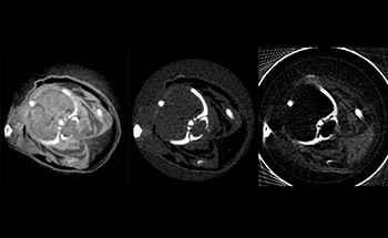
The researchers at the Technische Universität München (TUM) took multimodal scans of a formalin-preserved infant mouse that depicted its internal organs more clearly than standard imaging technology. In phase-contrast and dark-field-contrast images, the scientists could even distinguish between brown adipose tissue (the so-called “good” kind of body fat) and white adipose tissue, and they hope this ability could eventually lead to early diagnoses of tumors.
Ronald Ruth and Zhirong Huang of SLAC National Accelerator Laboratory (USA) developed the compact synchrotron based on inverse Compton scattering in 1998. The device consists of a 4.6-m-diameter ring that stores electrons at energies of 20 to 45 MeV before injecting them into an optical cavity, where they collide with an infrared laser pulse.
Traditional X-ray tubes emit photons at a wide range of energies, typically 10 to 100 keV, according to lead author Elena Eggl, a doctoral candidate at TUM. The compact synchrotron's X-ray beam has an intrinsic bandwidth of 3 percent, meaning its output is essentially monochromatic.
For the studies of the infant mouse, the team used a CCD detector with a spatial resolution of about 80 μm, Eggl says. CT scan resolution could be improved by using a detector with smaller pixels, at the cost of a higher radiation dose for the subject. A complete scan of the mouse took about 8 hours, but the exposure time will decrease as researchers continue to enhance the flux from the compact light source.
Eggl's group did the scans at the company in Palo Alto, Calif. (USA) that built the compact synchrotron. Once the instrument is installed at TUM, the researchers will conduct preclinical research on mammography, with human tumor samples from past surgeries, and on carbon composites and other non-living materials.