
Scatterings
Detecting Skin Cancer in Place
A Duke University researcher described how pump-probe imaging of melanin with near-infrared pulses allowed the researchers to image a developing melanoma directly on the skin.
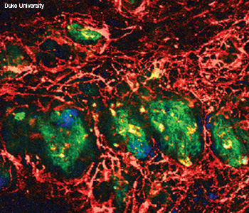 Imaging methods differentiate chemicals in living human skin.This false-color composite of near-IR pump-probe (blue and red) and second harmonic generation (green) imaging highlights hemoglobin and dermal collagen..
A multiphoton imaging technique called pump-probe spectroscopy can distinguish melanoma from benign lesions, thus providing pathologists with more information to determine whether tissue is cancerous. During the CLEO postdeadline papers session, Jesse W. Wilson of Duke University describes how pump-probe imaging of melanin with near-infrared pulses allowed the researchers to image a developing melanoma directly on the skin (CLEO Conference PDPB5, “Pump-Probe Melanoma Imaging: Applications to High-Resolution and In-Vivo Microscopy”).
…Log in or become a member to view the full text of this article.
This article may be available for purchase via the search at Optica Publishing Group.
Optica Members get the full text of Optics & Photonics News, plus a variety of other member benefits.