
Scatterings
Imaging Living Organisms with Sheets of Light
Caltech researchers developed a type of microscopy that simultaneously achieves high resolution, penetration depth and imaging speed, filling biologists’ need for fast 3-D imaging methods of organisms that don’t perturb the sample.
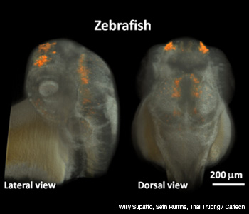 Three-dimensional live imaging of zebrafish showing both the embryo’s tissue (in white) and a fluorescent label driven by a gene-specific promoter (in orange).
Caltech researchers developed a type of microscopy that simultaneously achieves high resolution, penetration depth and imaging speed, filling biologists’ need for fast 3-D imaging methods of organisms that don’t perturb the sample. By combining two-photon excitation with light-sheet microscopy, Scott Fraser’s group demonstrated that the method provides deeper penetration than single-photon light-sheet microscopy. It also works much faster than point-scanning two-photon microscopy and doesn’t compromise normal biology (Nature Methods 8, 757; doi:10.1038/NMETH.1652).
…Log in or become a member to view the full text of this article.
This article may be available for purchase via the search at Optica Publishing Group.
Optica Members get the full text of Optics & Photonics News, plus a variety of other member benefits.