
Scatterings
Nanoscopy Uncovers Cells’ Secrets
Fluorescent optical microscopy is allowing researchers to image features much smaller than the diffraction limit of visible light.
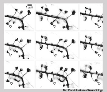 As the dendritic structures of neurons twist, the tips (indicated by arrows) turn cup-shaped. Images were obtained with stimulated emission depletion microscopy.
Fluorescent optical microscopy is allowing researchers to image features much smaller than the diffraction limit of visible light. This imaging technique, called nanoscopy, can yield resolutions down to hundredths of a micron—the size of large molecules.
…Log in or become a member to view the full text of this article.
This article may be available for purchase via the search at Optica Publishing Group.
Optica Members get the full text of Optics & Photonics News, plus a variety of other member benefits.