
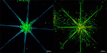
Left: the researchers patterned a star shape (shown here in blue) into their hydrogel structure. Right: after a few days, the cells (shown here in fluorescent green) migrated from the center of the star to outwards, following the pattern. [Image: TU Wien]
A team of researchers in Europe has shown how to use a laser beam to guide cells to the right spot in artificial tissues (Sci. Rep., doi: 10.1038/s41598-022-11612-y). The precise control of tissue properties afforded by the laser, the team believes, could advance “organ-on-a-chip” or “human-on-a-chip” technology—which could, in turn, allow scientists to study drugs’ effect on humans without animal testing.
Photochemical reactions
Scientists often use hydrogels—a special kind of cross-linked, water-logged polymer—to create artificial tissues, as hydrogels’ oxygen and nutrient permeabilities and mechanical characteristics are similar to human tissue. Once cells are embedded into hydrogels to create artificial tissues, however, it’s difficult to manipulate an individual cell’s “microenvironment” and make it travel within the hydrogel to the desired point. That makes creating new blood vessels in artificial tissues a challenge.
To tackle this issue, the research team behind the new work, based in Austria and Belgium, took advantage of multi-photon lithography, a high-resolution, light-based patterning technique already known for its ability to control cells’ microenvironments. The technique involves focusing a femtosecond laser beam at subdiffractional resolution onto a 3D target to start a photochemical reaction—which, properly controlled, can open up a channel in the hydrogel.
For their experiment, the researchers immersed their hydrogel into 4,4′-diazido-2,2′-stilbenedisulfonic acid (DSSA), a commercially available chemical that contains azido groups, for 24 hours. Then, they focused laser beams with 700 and 725 nm wavelengths and a scanning speed of 1000 mm/s to a predetermined pattern. The laser beams caused the azido groups consisting of three nitrogen atoms in DSSA molecules to photochemically decompose and form new bonds with the hydrogel, thereby softening the gel areas struck by the laser.
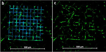
Left: a grid-like structure (blue) patterned into hydrogel. Right: after 13 days, the cells (green) aligned themselves along horizontal and vertical lines, following the pattern. [Image: S. Sayer et al. Sci. Rep. 12, 8626 (2022), CC BY 4.0]
After confirming their ability to pattern hydrogels using a laser, the researchers tested their method using human cells. First, the researchers patterned a mesh-like structure on hydrogel. When they observed human adipose-derived stem cells (hASCs) within the patterned hydrogel, the researchers found out that, after 13 days, 41% of the cells were aligned along vertical and horizontal lines, versus roughly 20% cells that aligned vertically or horizontally in the control sample. The researchers also observed desired cell migration when testing using a star-shaped pattern with hASCs and human umbilical vein endothelial cells.
Tissues on a chip
The researchers say that they expect their result to be useful when it comes to organ-on-a-chip or human-on-a-chip research, which involves growing artificial tissues placed on a chip. This type of research is useful when testing effects of nutrients or drugs on human cells, potentially eliminating the need for animal testing.
The new technique, according to study coauthor Tommaso Zandrini of Vienna University of Technology (TU Wien), Austria, now enables scientists to repeat human-on-a-chip experiments reliably, as the technique allows them to re-create several tissue samples with exactly the same microstructure. The researchers also expect their method to be useful for creating microvessels of blood in artificial tissues.