
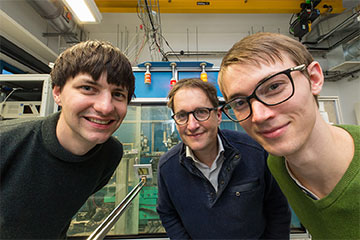
From left to right: Jakob Soltau, Tim Salditt and Paul Meyer in the laboratory where they carried out research into X-ray color imaging. Between Soltau and Salditt is the gold-coated plate used for their experiment. [Image: M. Osterhoff]
We’ve all seen the familiar black-and-white X-ray images of our own and others’ bodies. Sometimes, though, biomedical scientists need to distinguish between chemicals while performing advanced imaging techniques such as X-ray fluorescence (XRF).
Now researchers in Germany have proposed a method for quickly capturing full-field X-ray fluorescence images that show chemical concentrations and distributions in a target sample (Optica, doi: 10.1364/OPTICA.477809). The technique eliminates the need for time-consuming scanning of samples and could find uses in fields as diverse as industrial technology, materials science and art preservation.
Better focusing
In conventional XRF work, scientists scan a material with a focused beam and collect the photons emitted by the fluorescing substance. However, the X-ray sources used in typical laboratories produce incoherent radiation, which makes focusing the light a challenge—so users must resort to scanning an object with a narrow beam. Bright synchrotron sources have more coherence but require travel to a specialized facility.
In recent years, some scientists have tried full-field XRF imaging with coded aperture masks: grids or gratings that cast a known shadow of high-energy radiation on the image plane. When combined with analog image reconstruction, a type of mask called a modified uniformly redundant array (MURA) could perform the job, but small imperfections in its position could ruin the experiment.
Fresnel zone plate meets algorithm
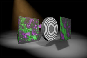
Artistic representation showing the creation of an image using the newly developed method. Two colors—green and magenta—are emitted by fluorescing atoms in the sample (left) due to X-ray excitation. The round, gray object represents an optic casting a shadow on the detector. The algorithm then produces an actual image with two colors, the intensity of which represents the density of the fluorescing atoms within the sample. [Imaget: M. Osterhoff]
To achieve the level of focus required for full-field imaging in the laboratory, researchers at the University of Göttingen, Germany, devised a Fresnel zone plate (FZP) that could be inserted between the fluorescence-emitting sample and the detector. Whereas the structure and position of a MURA must be precisely known, scientists can use fast algorithms to reconstruct the data from the zone plate setup.
In the Göttingen team’s experiment, an X-ray tube with a rotating anode emitted incoherent rays, subsequently collimated by a pair of curved mirrors. The collimated beam hit the target sample at an angle of 10°. The resulting fluorescence headed in all directions, with part of it passing through the gold-coated zone plate and hitting an X-ray color camera. The camera distinguished between detected photons with different energies, and computational algorithms completed the analysis. “We have developed an algorithm that allows us to quickly and robustly create a sharp image simultaneously for each X-ray color," explains author Jakob Soltau in a press release.
According to Tim Salditt, a Göttingen professor who led the research team, the main challenge in the experiment was “finding the sweet spot” that balanced all experimental parameters, such as field of view, photon flux, fluorescence yield and geometric constraints.
Many potential applications
Salditt says his research group is very interested in 3D imaging of biological tissues. Optimizing the new full-field XRF technique would allow scientists to image the structure and local chemical composition of a sample in a single scan. XRF is also used to examine the quality of manufactured computer chips and to analyze soil samples.
In a few months, the Göttingen scientists will use their scheduled beamtime on a synchrotron source to improve their experimental parameters for higher resolution and shorter acquisition times, Salditt says.