
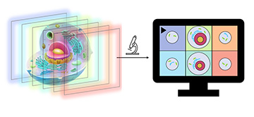
The multifocus microscope provides clear images by using light sheets to scan the planes of a specimen. [Image: UiT]
Researchers in Norway have developed and tested a multifocus microscopy prototype that reportedly can improve axial resolution and eliminate background haze in images of subcellular structures in dense tissues (Optica, doi: 10.1364/OPTICA.468583). The microscope—which does its magic via oblique optical sectioning—captures a speedy 35 volumes per second with less light than traditional microscopes, making it less damaging to photosensitive live tissues and fluorescent markers.
Florian Ströhl and his colleagues at UiT The Arctic University of Norway and the University Hospital of North Norway have applied for a patent for their device and are looking for industrial partners to help bring the multifocus microscope to market.
Speeding up imaging with optical sectioning
Common methods for 3D microscopy bathe a specimen in light while capturing a series of pixels that must be stitched together to form an image. This approach works well for static objects but is not practical for living and moving samples.
Ströhl and his colleagues came up with a scalable 3D microscopy method that uses optical sectioning to improve image capture speed and axial resolution. It also requires less light exposure than traditional methods, which may protect live cells that are prone to photodamage and cause less photobleaching in fluorescent labels.
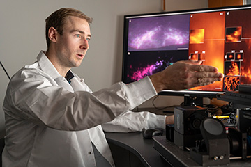
Study first author Florian Ströhl, UiT The Arctic University of Norway. [Image: K. Rydland/UiT]
The team named its imaging approach SOLIS—short for scanned oblique light-sheet instant-volume sectioning. SOLIS uses a dithered oblique light-sheet to scan the planes of a specimen during a single exposure, which increases axial resolution and reduces light exposure. Fluorescence from each plane is then mapped onto a camera chip with a multifocus optical element—such as a prism, a grating and a beam-splitter cascade—that generates displaced images of corresponding image planes. Adjusting the multifocal optical element enables the mapping of tilted illumination planes onto single lines on the camera, while a synchronized rolling-shutter and light-sheet readout mode are used for optical sectioning.
Theoretical verification and prototype demonstration
To characterize SOLIS’ performance, the researchers simulated widefield 3D microscopy with a scanned light sheet and SOLIS in two modes: with an on-axis propagating light sheet and with an oblique light sheet. They then compared the widefield simulation with the two SOLIS ones. Results of this virtual imaging demonstration showed that both SOLIS models did indeed eliminate background haze. The SOLIS simulations were also able to image a 6.4-µl cube containing randomly distributed point-emitters at resolutions in agreement with theoretical calculations and close to the simulated widefield microscope’s performance.
Next, the team used a SOLIS prototype with a multifocus optical beam-splitter cascade to record crisp images of endothelial cell monolayers at a volumetric frame rate of 35 Hz, while moving the imaging stage at a speed of 35 µm/s. The researchers also demonstrated subcellular-scale volumetric imaging with a cube of live cultured heart cells. Using SOLIS, they revealed the distribution of mitochondria and mitochondria-derived vesicles at depths up to 30 µm.
The research team is now working on upgrading its design to create a multifocus microscope that is easier to use. In the meantime, the researchers are making the prototype available to local partners.
